The conscious and subconscious mind crave orderliness and organization, with each item of data clearly recorded in multisensory detail, and filed in its most appropriate and accessible place. This is why we have routines, rituals and recurring cycles of thought (which sometimes haunt us and which sometimes help us). Look at the picture above. At first, it appears as a meaningless bunch of letters -- but as we study it consciously, it becomes a plainly-worded statement of fact. In fact, when we look at incomplete pictures or have "blind spots" in our foveal or peripheral vision, our minds tend to fill in those blanks for us. This effect is responsible for many optical illusions. And what we see (or think that we are seeing) is a significant input into what we will be thinking. Peripheral vision plays more tricks on us than foveal vision, but our foveal vision can be made to play tricks on us as well.
Let's get ourselves some definitions of these two types of vision from Wikipedia, the source for just about anything...
Foveal
From Wikipedia, the free encyclopedia

Schematic diagram of the human eye, with the fovea at the bottom.
The foveal system of the human eye is the only part of the retina that permits 100% visual acuity.
The line-of-sight is a virtual line connecting the fovea with a fixation point in the outside world.
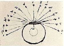
The discovery of the line-of-sight is attributed to Leonardo da Vinci.1
His main experimental finding was that there is only a distinct and clear vision at the line-of-sight, the optical line that ends at the fovea. Although he did not use these words literally he actually is the father of the modern distinction between foveal vision (a more precise term for central vision) and peripheral vision.

Leonardo da Vinci: The eye has a central line and everything that
reaches the eye through this central line can be seen distinctly.
Leonardo da Vinci, (1452-1519) was the first person known in Europe to recognize the special optical qualities of the eye. He derived his insights partly through introspection but mainly through a process that could be described as optical modelling. Based on dissection of the human eye he made experiments with water-filled crystal balls. He wrote "The function of the human eye, ... was described by a large number of authors in a certain way. But I found it to be completely different."
---------------
Peripheral vision is a part of vision that occurs outside the very center of gaze. There is a broad set of non-central points in the field of view
that is included in the notion of peripheral vision. "Far peripheral"
vision exists at the edges of the field of view, "mid-peripheral" vision
exists in the middle of the field of view, and "near-peripheral",
sometimes referred to as "para-central" vision, exists adjacent to the
center of gaze.
In vision-related fields such as physiology, ophthalmology, or optometry, the inner boundaries of peripheral vision are defined more narrowly in terms of one of several anatomical regions of the central retina, generally the fovea.
The fovea is a cone-shaped depression in the central retina measuring 1.5 mm in diameter, corresponding to 5° of the field of vision.The outer boundaries of the fovea are visible under a microscope, or with microscopic imaging technology such as OCT or microscopic MRI. When viewed through the pupil, as in an eye exam (using ophthalmoscope or retinal photography) only the central portion of the fovea is visible. Anatomists refer to this as the clinical fovea, and say that it corresponds to the anatomical foveola, a structure with a diameter of 0.35 mm corresponding to 1 degree of the field of vision. In clinical usage the central part of the fovea is typically referred to simply as the fovea.
In terms of visual acuity, "foveal vision" may be defined as the part of the retina in which visual acuity is at least 20/20 (6/3 metric). This corresponds to the foveal avascular zone (FAZ) with a diameter of 0.5 mm representing 1.5° of the visual field. Although often idealized as perfect circles, the central structures of the retina tend to be irregular ovals. Thus, foveal vision may also be defined as the central 1.5-2° of the field of vision. Vision within the fovea is generally called central vision, while vision outside of the fovea is called peripheral vision.
A ring-shaped region surrounding the fovea, known as the parafovea, is sometimes taken to represent an intermediate form of vision called paracentral vision. The parafovea has an outer diameter of 2.5 mm representing 8° of the field of vision. The macula, a region of the retina defined as having at least two layers of (bundles of nerves and neurons) is sometimes taken as defining the boundaries of central vs. peripheral vision. The macula has a diameter of 5.5 mm and corresponds to 18° of the field of vision. When viewed from the pupil, as in an eye example, only the central portion of the macula is visible. Known to anatomists as the clinical macula (and in clinical setting as simply the macula) this inner region is thought to correspond to the anatomical fovea.
The dividing line between near and mid peripheral vision at 30° radius is based on several features of visual performance. Visual acuity declines by about 50% every 2.5° from the center up to 30°, at which point the decline in visual acuity declines more steeply. Color perception is strong at 20° but weak at 40° . 30° is thus taken as the dividing line between adequate and poor color perception. In dark-adapted vision, light sensitivity corresponds to rod density, which peaks just at 18° . From 18° towards the center rod density declines rapidly. From 18° away from the center, rod density declines more gradually, in a curve with distinct inflection points resulting in two humps. The outer edge of the second hump is at about 30° , and corresponds to the outer edge of good night vision.
Peripheral vision is weak in humans, especially at distinguishing color and shape. This is because receptor cells on the retina are greater at the center and lowest at the edges (see visual system for an explanation of these concepts). In addition, there are two types of receptor cells, rod cells and cone cells; rod cells are unable to distinguish color and are predominant at the periphery, while cone cells are concentrated mostly in the center of the retina, the fovea.
Flicker fusion threshold is higher for peripheral than foveal vision. Peripheral vision is good at detecting motion (a feature of rod cells).
Central vision is relatively weak at night or in the dark, when the lack of color cues and lighting makes cone cells far less useful. Rod cells, which are concentrated further away from the retina, operate better than cone cells in low light. This makes peripheral vision useful for seeing movement at night. In fact, pilots are taught to use peripheral vision to scan for aircraft at night.
The distinctions between foveal (sometimes also called central) and peripheral vision are reflected in subtle physiological and anatomical differences in the visual cortex. Different visual areas contribute to the processing of visual information coming from different parts of the visual field, and a complex of visual areas located along the banks of the interhemispheric fissure (a deep groove that separates the two brain hemispheres) has been linked to peripheral vision. It has been suggested that these areas are important for fast reactions to visual stimuli in the periphery, and monitoring body position relative to gravity.
Peripheral vision can be practiced; for example, jugglers that regularly locate and catch objects in their peripheral vision have improved abilities. Jugglers focus on a defined point in mid-air, so almost all of the information necessary for successful catches is perceived in the near-peripheral region.
Boundaries
Inner boundaries
The inner boundaries of peripheral vision can be defined in any of several ways depending on the context. In common usage or everyday language the term "peripheral vision" is commonly used to refer to what in technical usage would be called "far peripheral vision." This is vision outside of the range of stereoscopic vision. It can be conceived as bounded at the center by a circle 60° in radius or 120° in diameter, centered around the fixation point, i.e., the point at which one's gaze is directed.1 In common usage, peripheral vision may also refer to the area technically known as "mid peripheral vision," defined by a circle 30° in radius or 60° in diameter.In vision-related fields such as physiology, ophthalmology, or optometry, the inner boundaries of peripheral vision are defined more narrowly in terms of one of several anatomical regions of the central retina, generally the fovea.
The fovea is a cone-shaped depression in the central retina measuring 1.5 mm in diameter, corresponding to 5° of the field of vision.The outer boundaries of the fovea are visible under a microscope, or with microscopic imaging technology such as OCT or microscopic MRI. When viewed through the pupil, as in an eye exam (using ophthalmoscope or retinal photography) only the central portion of the fovea is visible. Anatomists refer to this as the clinical fovea, and say that it corresponds to the anatomical foveola, a structure with a diameter of 0.35 mm corresponding to 1 degree of the field of vision. In clinical usage the central part of the fovea is typically referred to simply as the fovea.
In terms of visual acuity, "foveal vision" may be defined as the part of the retina in which visual acuity is at least 20/20 (6/3 metric). This corresponds to the foveal avascular zone (FAZ) with a diameter of 0.5 mm representing 1.5° of the visual field. Although often idealized as perfect circles, the central structures of the retina tend to be irregular ovals. Thus, foveal vision may also be defined as the central 1.5-2° of the field of vision. Vision within the fovea is generally called central vision, while vision outside of the fovea is called peripheral vision.
A ring-shaped region surrounding the fovea, known as the parafovea, is sometimes taken to represent an intermediate form of vision called paracentral vision. The parafovea has an outer diameter of 2.5 mm representing 8° of the field of vision. The macula, a region of the retina defined as having at least two layers of (bundles of nerves and neurons) is sometimes taken as defining the boundaries of central vs. peripheral vision. The macula has a diameter of 5.5 mm and corresponds to 18° of the field of vision. When viewed from the pupil, as in an eye example, only the central portion of the macula is visible. Known to anatomists as the clinical macula (and in clinical setting as simply the macula) this inner region is thought to correspond to the anatomical fovea.
The dividing line between near and mid peripheral vision at 30° radius is based on several features of visual performance. Visual acuity declines by about 50% every 2.5° from the center up to 30°, at which point the decline in visual acuity declines more steeply. Color perception is strong at 20° but weak at 40° . 30° is thus taken as the dividing line between adequate and poor color perception. In dark-adapted vision, light sensitivity corresponds to rod density, which peaks just at 18° . From 18° towards the center rod density declines rapidly. From 18° away from the center, rod density declines more gradually, in a curve with distinct inflection points resulting in two humps. The outer edge of the second hump is at about 30° , and corresponds to the outer edge of good night vision.
Outer boundaries
The outer boundaries of peripheral vision correspond to the boundaries of the visual field as a whole. For a single eye, the extent of the visual field can be defined in terms of four angles, each measured from the fixation point, i.e., the point at which one's gaze is directed. These angles, representing four cardinal directions, are 60° superior (up), 60° nasal (towards the nose), 70-75° inferior (down), and 100-110° temporal (away from the nose and towards the temple). For both eyes the combined visual field is 130-135° vertical and 200-220° horizontal.Characteristics
The loss of peripheral vision while retaining central vision is known as tunnel vision, and the loss of central vision while retaining peripheral vision is known as central scotoma.Peripheral vision is weak in humans, especially at distinguishing color and shape. This is because receptor cells on the retina are greater at the center and lowest at the edges (see visual system for an explanation of these concepts). In addition, there are two types of receptor cells, rod cells and cone cells; rod cells are unable to distinguish color and are predominant at the periphery, while cone cells are concentrated mostly in the center of the retina, the fovea.
Flicker fusion threshold is higher for peripheral than foveal vision. Peripheral vision is good at detecting motion (a feature of rod cells).
Central vision is relatively weak at night or in the dark, when the lack of color cues and lighting makes cone cells far less useful. Rod cells, which are concentrated further away from the retina, operate better than cone cells in low light. This makes peripheral vision useful for seeing movement at night. In fact, pilots are taught to use peripheral vision to scan for aircraft at night.
The distinctions between foveal (sometimes also called central) and peripheral vision are reflected in subtle physiological and anatomical differences in the visual cortex. Different visual areas contribute to the processing of visual information coming from different parts of the visual field, and a complex of visual areas located along the banks of the interhemispheric fissure (a deep groove that separates the two brain hemispheres) has been linked to peripheral vision. It has been suggested that these areas are important for fast reactions to visual stimuli in the periphery, and monitoring body position relative to gravity.
Peripheral vision can be practiced; for example, jugglers that regularly locate and catch objects in their peripheral vision have improved abilities. Jugglers focus on a defined point in mid-air, so almost all of the information necessary for successful catches is perceived in the near-peripheral region.
Functions
The main functions of peripheral vision are:- recognition of well-known structures and forms with no need to focus by the foveal line of sight,
- identification of similar forms and movements (Gestalt psychology laws),
- delivery of sensations which form the background of detailed visual perception.
Both foveal and peripheral vision can be strengthened just through frequent use and exercise. These exercises are worthwhile because they assist us in seeing a true picture of the world around us with minimal "filling-in" by the ever-orderly imagination. If our minds are provided with greater amounts of higher resolution "real" visual input, this can only serve us better. Practice those eye exercises!
Each of the links below my signature leads to an interesting optical illusion. Take a look at each, and note how your mind completes incomplete patterns and creates the illusion of motion when objects being observed are actually still.
Enjoy the experience.
Douglas E. Castle for Braintenance
http://www.michaelbach.de/ot/
http://kids.niehs.nih.gov/games/illusions/
http://www.brainbashers.com/opticalillusions.asp


BRAINTENANCE: Train, Strain And Improve Your Brain. Expand Your Mind.
http://braintenance.blogspot.com
Braintenance contains articles, resources, exercises, games and specially-designed protocols to improve the power of your brain and your mind in every significant aspect, including memory, cognition, IQ, plasticity, creativity and problem-solving ability.
Key Terms: brain, mind, cognitive enhancement, memory, brain gym exercises, IQ, plasticity, mind expansion, creativity, meditation, altered states, perception, self-hypnosis, self-growth, neuron, artificial intelligence, learning, somatic intellect, mathematics, language, dissonance, individualism, herd mentality, puns and word games, linguistics, genius, emotion, subconscious, unconscious, intuition, instinct, psychedelic, reality, learning curve, probability, collective consciousness


No comments:
Post a Comment